Researchers use 3D bioprinted tumor models to transform cancer treatment
The SDSU Tambasco Lab’s lifelike models work to improve radiation treatment while mentoring students in cutting-edge medical physics.
In associate professor Mauro Tambasco’s lab, researchers at all levels work together to develop innovative approaches to fighting cancer.
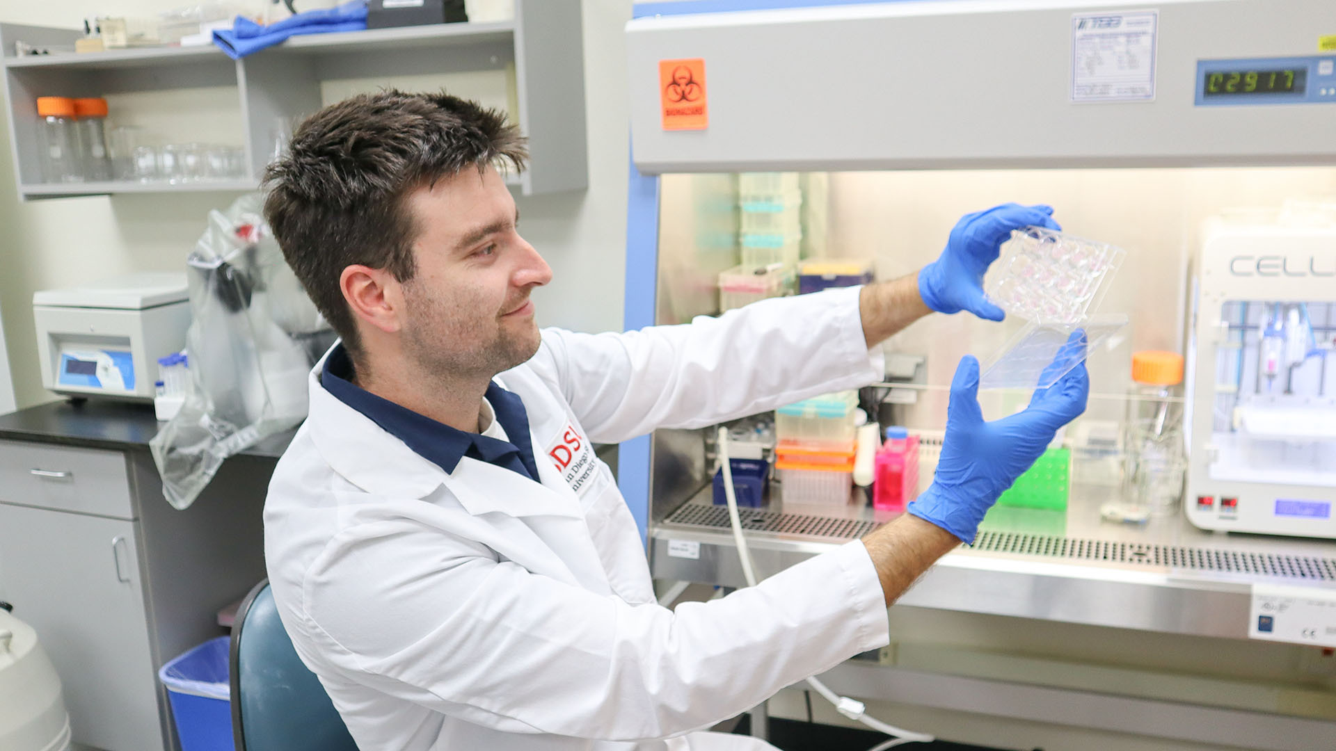
By creating 3D models of tumors, the lab can better study the intricacies of these cancerous clumps of cells. Researchers form the tumor models with cutting-edge bioprinting technology, which produces standardized models with the properties and functions of real cells, allowing for a more realistic and accurate way to study and test treatments.
“A lot of radiotherapy is based off two-dimensional cell models, which don't capture a lot of the immune and vascular effects that a tumor model would,” said Harry Glazebrook, medical physics master’s student in the College of Sciences.
“3D bioprinting allows you to mimic those cell to cell interactions." Glazebrook added. "You can print a whole tumor with vasculature, perfusion, blood flow, things that really only exist inside the human body. Then you could take that model and test different radiotherapy or chemical techniques on it to make sure we're fighting cancer as effectively as possible.”
The two-dimensional models currently used to determine cancer patients’ radiation doses and types do not provide this comprehensive perspective. Using lifelike 3D models, the Tambasco lab works to fine-tune radiation therapy, making it more precise and efficient.
For example, while many patients are prescribed small, uniform doses of radiation on a daily basis over several weeks, the Tambasco lab uses its models to test whether higher doses delivered only to specific parts of the tumor could trigger an immune response with the potential to improve patient survival.
“3D bioprinting has been used before but mostly with chemotherapy drugs,” Glazebrook said. “We're trying to translate that technology so we can make radiotherapy as effective as possible.”
Beyond the science
From writing workshops to research experiences, Glazebrook said SDSU’s graduate support programs have helped prepare him for a career as a health professional.
“As a medical physicist, I will be working in the clinic with radiation therapy and helping treat people with cancer,” he said. “This research is really beneficial because it gives me a closer perspective on the biology of cancer, how it works and how I can better help people.”
In turn, Glazebrook has served as a mentor to undergraduate students in the lab, helping them develop varied technical and communication skills he said lets them “leave this lab with a lot of skills they wouldn't have otherwise.”
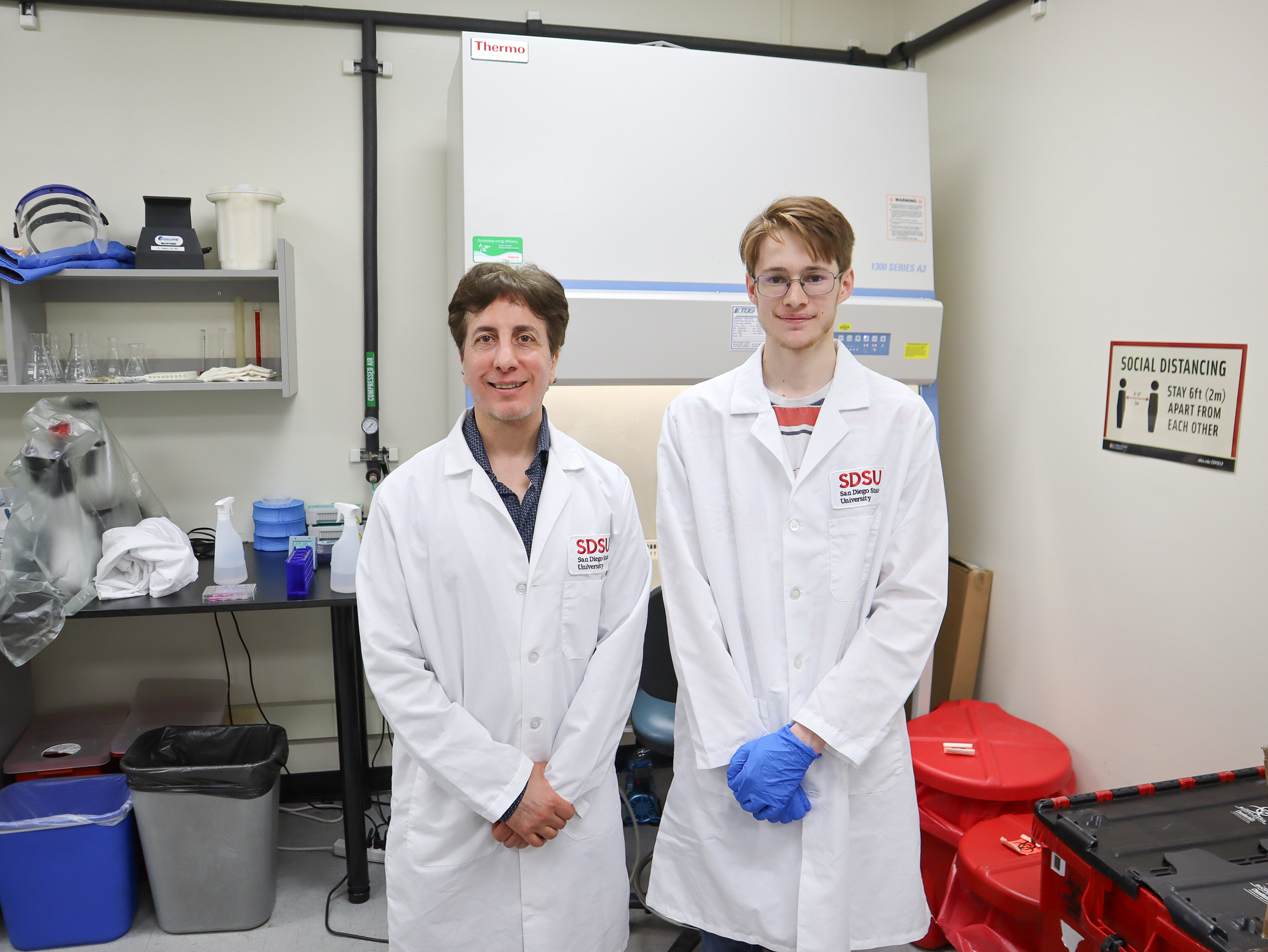
One of these students is Connor Schmidt, a fourth-year student whose project is supported by the SDSU Undergraduate Research, Scholarship, and Creative Activities Program (SURP). In the lab, Schmidt examines how different levels of radiation affect the elasticity and viscosity of the tumor models. Scientists can use this insight to understand the role these biomechanical properties play in tumor regression and predict these effects in real tumors.
“I've always been interested in how I can apply physics and science to real things and real people,” Schmidt said.
As a physics student, Schmidt embraced the challenge of learning the biological concepts and techniques central to medical physics research. Through hands-on experience and by seeing the real-world impact of his work in the Tambasco Lab, he developed a strong passion for the field and now plans to pursue graduate studies to continue his training.
“As I was researching possible careers I could go into with a physics degree, medical physics really stood out to me as something where I would be actively working in a hospital and helping develop better treatments for cancer,” Schmidt said.
For more information on medical physics at SDSU, visit the program website.